The opacity of the human form has posed a continuing challenge to medical science – obscuring the efforts of diagnostic medicine. This has motivated a recent UCLA study that proposes to harness cutting-edge 3D visualization techniques to enhance prostate cancer detection by combining real-time ultrasound with multi-parametric MRI. Thus, the traditional challenge of detecting prostate cancer using antigen blood tests that yield unclear results, could well become a thing of the past. Fusing Modalities In terms of imaging modality, ultrasound has always been considered a cost-efficient diagnosis methodology. However, image contrast is an issue, and the output is far inferior to CT scans. The Journal of Ultrasound Medicine published a report comparing CT-guided hepatic lesion biopsy to ultrasound fusion technology, and the results are telling. In terms of diagnostic yield, ultrasound fusion outperformed the CT-guided method by a margin of 2.1%. The mean procedure duration also varied substantially, with the fusion technique taking 30 minutes less than what the CT scan took to perform. Although ultrasound fusion has allowed urologists to transition from blind, systematic biopsies to a more streamlined, targeted approach, it has its share of drawbacks. When it comes to the field of musculoskeletal diagnostics, the method generally relies on conventional grayscale (or B-mode) ultrasound imaging. While the Doppler principle has proved itself in evaluating cardiac muscle function, there is much to be desired in terms of its ability to measure strain, strain rate, and tissue velocities. To that effect, researchers are already exploring the idea of building an ultrasound platform that leverages high-frequency transducers to produce a tighter beam of greater efficiency in terms of maintaining outstanding spatial and contrast resolution. It will allow physicians to image superficial musculoskeletal structures with much greater accuracy.
However, the jury is still out on the most effective modality for musculoskeletal imaging. Given the complexity of the human anatomy, no single modality is optimal, and must be based on the area or condition that needs to be diagnosed. For example, detecting disc pathology and chord abnormalities is only possible through MRI since ultrasound has been reported to have 70% or less accuracy in detecting lumbar disc herniation. At the other end of the spectrum, ultrasound scan (USS) is a far more effective solution to dynamically assess subacromial subdeltoid bursa and rotator cuffs, while highlighting active impingement. Although modality differences may be attuned to address specific diagnostic requirements, the case for better musculoskeletal imaging can be furthered by synergizing the two imaging techniques. Ultrasound guided magnetic resonance arthography has become an effective modality for imaging hands, lower limbs, hip joints, flexor, extensor and peroneal tendons. Now that we have come closer to identifying the appropriate diagnostic imaging solution, we can accurately identify the underlying condition of the patient. But, how exactly do you address a complex musculoskeletal injury like a fractured lumbar, and maximize patient outcome success rate?
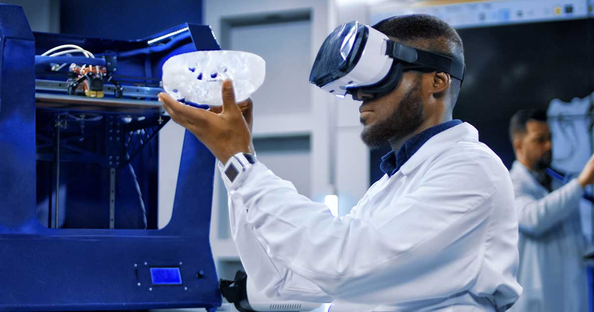
Digital engineering does not just facilitate better diagnosis, but also helps develop solutions – 3D printed models and implants. The Children’s Hospital of Philadelphia is currently conducting research into using a combination of low dose X-ray, MRI, ultrasound and CT scans to develop precise digital 3D models of bones. In combination with gait and motion analysis tools, it allows physicians to gain a 360-degree view of the spine, and even simulate how the bones and joints move together while the patient is in motion. Expanding on the scope of this initiative, the research group plans to use virtual models to print 3D replicas of the spine and the ribcage, enabling physicians to visualize and plan for delicate surgeries. Digital imaging assets can be utilized to facilitate even greater advances in the field of musculoskeletal medicine. Current technology already allows us to craft biomedical implants for cranial and facial replacements. A leading high performance additive manufacturer was recently granted a patent by European authorities for its proprietary manufacturing process, which creates 3D printed, customized implants for bone replacement. At the confluence of biomedical sciences, imaging and 3D printing technology, stem cell research may hold the key to revolutionize replacement implants. Scientists at Canada’s Mount Sinai Centre for Regenerative Medicine and Musculoskeletal Research, and the University of Rochester Medical Center are furthering the cause, testing ways to use regenerated tissue and 3D printing to create the next generation of biodegradable implants.


